Advanced Mapping Offers New Insights
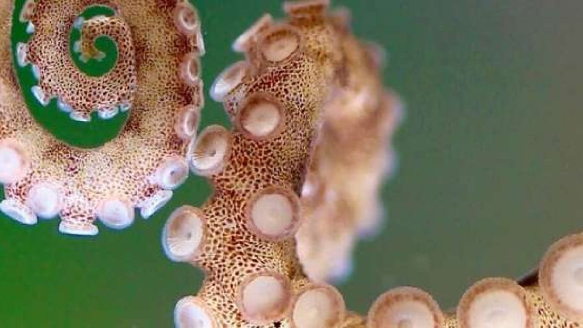
The research, led by Robyn Crook, Associate Professor and Associate Chair of the SF State Biology Department, addresses a long-standing query in marine biology: how do octopus arms handle such complicated behaviours with out fixed enter from the mind? Using superior 3D imaging methods, Gabrielle Winters-Bostwick, a postdoctoral fellow, and Diana Neacsu, a graduate pupil, have created detailed anatomical and molecular maps that reveal the distinctive organisation of octopus arms.
Winters-Bostwick’s examine used molecular tags to focus on various kinds of neurons, unveiling that the neurons on the arm’s tip differ considerably from these situated close to the central mind. Meanwhile, Neacsu employed 3D electron microscopy to discover the structural organisation, discovering repeating patterns in nerve branches and ganglia throughout the arm.
Crucial Role of Advanced Imaging
The research had been made attainable by SF State’s superior imaging expertise, notably the Leica STELLARIS microscope housed within the University’s Cellular and Molecular Imaging Centre (CMIC). This useful resource has been a game-changer for the crew. “Without this microscope, a lot of our analysis would not have been attainable,” Crook remarked.
The findings from these maps may revolutionise our understanding of octopus physiology, and the instruments developed will possible be adopted by different labs learning cephalopod neuroscience. Researchers purpose to analyze how octopus arms reply to stimuli and discover the evolutionary causes behind their distinctive nervous system construction.


